-
Achromatic lens: a lens
that is specially designed and coated to correct for
the tendency of light to separate into colors when
passing through glass. An achromatic lens corrects
this such that colors are more accurate after being
magnified.
Top
-
Binocular microscope:
A compound microscope with two eyepieces viewing down
a single optical channel and objective. This is different
than a stereo microscope, which
has a separate optical channel for each eye.
Top
-
C-mount & CS-mount:
Also referred to as C/CS-mount, it is a threaded standard
developed for mounting a lens to a camera. The mount
is most commonly used for video cameras (i.e., CCTV
cameras, not camcorders), and is used to mount cameras
to microscopes. The mechanical definition of both
standards is 1" diameter, 32 TPI (threads per inch),
male on the lens (or microscope) side and female on
the camera side. The optical definition of the C-mount
is that the image reaches the focal plane, or camera's
detector, at 17.5mm past the edge of the lens' (or
microscope's) mounting threads. The CS-mount is identical
in all respects except the focal plane is 12.5mm past
the mounting threads. A CS-mount camera can be mounted
on a C-mount lens or microscope by using a 5mm extension
ring. See also: T-mount.
Top
-
Coaxial Controls: A configuration
where one knob is centered on top of another. For
example, coarse and fine focus may have a larger coarse
focus knob with a fine focus knob on top of it (so
the center of both knobs is on the same axis). Also
commonly used for Mechanical
Stage X/Y movement knobs.
Top
-
Compound microscope: A
microscope with multiple lenses, however this definition
describes virtually all modern microscopes. It would
typically include multiple user-selectable objective
lenses of varying magnifications and present a two-dimensional
view. Also see: stereo microscope.
Top
-
Condenser: The light
rays from the illuminator are condensed and focused
through this lens in the center of the stage, providing
better image resolution.
Top
-
Digital microscope: A microscope
and video camera combination with a digital output
such as USB or firewire. The microscope often includes
software to display the image on a PC.
Top
-
DIN Standard Objectives: (Deutsches
Institut fuer Normung) An international standard which
dictates the design compatibility of the objective
lens. DIN standard objectives from one manufacturer
can be used in another manufacturer's DIN standard
compatible microscope.
Top
-
Doublet lens: a lens design
that is actually two different lenses cemented together
(usually one positive magnifier and one negative).
This design is used in widefield
eyepieces to obtain significantly better color
performance than single lens designs.
Top
-
Dual-view microscope:
A monocular microscope with
a second, vertical viewing port. The vertical port
can be used with an eyepiece for a second person,
such as an instructor, to view the specimen, or it
can be used with an adapter and a video or still camera.
See also: trinocular microscope.
Top
-
Eyepiece or Ocular: The
lens closest to your eye when looking through a microscope.
A binocular or stereo microscope will have two, a
monocular microscope will have one. The lensalso plays
a critical role in the total system magnification.
See also widefield eyepiece.
Top
-
Eyepiece Tube or Eyetube:
The tube into which the eyepiece lens (ocular) is
set. This is usually presented at an angle for comfortable
viewing. It may also be mounted in a vertical position
such as on a trinocular or dual-view microscope for
either a second viewer, or for a camera designed to
fit inside an eyetube.
Top
-
FPS: frames per second: Used
to indicate the speed in which a video image is refreshed
and displayed on a monitor. In video microscopy this
is usually controlled by the camera. The faster the
refresh rate (number is larger), the "smoother" any
movement of the specimen will appear.
Top
-
Interpupillary Distance:
Distance between the two eyepieces. Typically it is
adjustable to accommodate different users. Some microscopes
also have graduated scales to indicate the actual
distance between the eyepieces, allowing a user to
determine the optimum number and then quickly set
it before each use.
Top
-
Koehler Illumination: A
highly effective illumination design.
Top
-
Magnification: Multiply
the magnification of the eyepiece by the magnification
the objective lens for the total magnification at
that power. 400x or 1000x is necessary for studying
cells and cell structure.
Top
-
Mechanical Stage: A mechanism
mounted on top, or as part, of the stage that allows
the operator to move the specimen slide in the X or
Y direction by turning a knob. Very useful at higher
magnifications as it can be difficult to move the
slide by hand. Most mechanical stages come with a
graduated scale so you can see how far the slide has
been moved or keep track of the position of various
objects on the slide.
Top
-
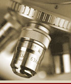
Objective lens: The lens
in a microscope closest to the specimen. In a compound
microscope there are usually 3, 4 or 5 objective lenses
allowing a selection of magnification levels.
Top
-
Oil Immersion lens: A lens designed
to be immersed in oil. A drop of immersion oil is
placed on top of the cover glass and the lens is slowly
lowered until it rests in the oil. This allows the
light to pass through oil rather than air, and at
higher magnifications results in a crisper, higher
contrast image. Primarily seen on more advanced systems.
Top
-
Parcentered: A lens
design such that specimens that appear centered in
the field of view at one magnification level will
also appear centered when the magnification level
is changed. See also: parfocal
Top
-
Parfocal: A lens
design such that specimens that appear in focus at
one magnification will also appear focused when the
magnification level is changed. The depth of field
(how much of a specimen's height will appear in focus
at one time) changes significantly when magnification
is changed. The higher the magnification, the shallower
the depth of field. See also: parcentered
Top
-
Phase Contrast: A technique
using special objectives and condenser system to enhance
the contrast of unstained, relatively transparent
specimens such as blood and other tissue cells, thereby
allowing microscopic viewing of living tissue. It
is a sophisticated technique that shifts the light
"phase" 1/4 wavelength, causing any light deviated
by the specimen to appear dark on a light background.
Top
-
Rack Stop: A safety feature
consisting of a mechanical stop, typically adjustable,
which prevents the objective
lens from hitting the microscope stage.
Top
-
Seidentopf: a head design
where the interpupillary adjustment (increasing or
decreasing the distance between the eyepieces) is
achieved by twisting the eyepieces in an up and down
arc motion like binoculars.
Top
- Slip
Clutch: A safety device usually located on the
focus knob allowing the knob to "slip" and continue
turning when it reaches the end of its travel, or
if it runs into the stage. Due to the gear ratios
involved, without this it may be possible to damage
the mechanism by applying too much force to the knob
after it has reached the end.
Top
- Stage:
The platform that holds the slide up beneath the
objective lens.
Top
-
Stereo microscope a.k.a. dissecting
microscope: A microscope with a separate optical
channel for each eye (eyepieces and objectives) which
allows viewing in three dimensions. See also: compound
microscope.
Top
- Turret
or Objective Turret: The rotatable metal piece
into which the microscope's objective
lenses are attached. A "turret" style stereo
microscope refers to the type that has more than
one objective lens which can then be rotated into
position. On a compound microscope
the turret is the ring holding the objective lenses
allowing the operator to rotate them into position
as needed.
Top
-
T-mount: A photographic
mechanical mounting standard developed in 1957 originally
intended as a universal lens mount for 35mm cameras.
There are now T-mounts available for a large variety
of digital and film cameras making it a good method
for mounting cameras to microscopes. The thread (a.k.a.
T-thread) is specified as 42mm diameter and 0.75mm
pitch, or M42-.75. See also: C-mount
Top
-
Trinocular microscope:
A binocular microscope with
a third, vertical viewing port. The vertical port
can be used with an eyepiece for a second person,
such as an instructor, to view the specimen, or it
can be used with an adapter and a video or still camera.
Click here
for an example. See also: dual-view
microscope.
Top
-
Widefield eyepiece (WF): an
eyepiece with an achromatic
doublet lens designed in such
a way that itdoes not have to be limited to viewing
only in its center, and the portion of the lens that
allows non-distorted viewing is larger than a normal
lens. This appears to the user as a bigger aperture
or "hole" to look through. It therefore has the advantage
of being easier to use and more forgiving of a user's
head movements. An eyepiece listed as WF10X/18mm would
indicate it has a widefield achromatic doublet lens,
10X magnification and is 18mm in diameter.
Top
|